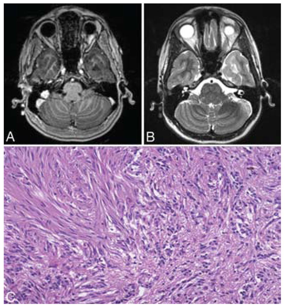
FIG. 4. Case 3.
A: Axial T1-weighted MR image with contrast showing a nonenhancing lesion in the left anterior temporal lobe. B: Axial T2-weighted MR image obtained at the same level, showing the hyperintense tumor signal. C: Photomicrograph displaying small clusters of monomorphous cells in the right side of the panel in opposition to more densely packed fascicles of elongated tumor cells in the left side of the panel. This pattern, which is reminiscent of a schwannoma, frequently can be seen in these lesions. This pattern may be a source of diagnostic confusion given the size of the specimen. H & E, original magnification × 64.

