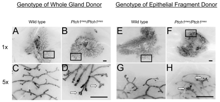
Fig. 3.
Whole gland and epithelial fragment transplantation into Rag1-/- hosts. The genotype of the epithelial fragment donor is shown above each column, and the magnification values of these inverted fluorescence images to the left. The boxed regions in A,B,E,F are shown at higher magnification in C,D,G,H. (A,C) Wild-type epithelial fragment showing complete filling of the available fat pad and normal duct morphology (A), with blunt or rounded duct termini (C). (B,D) Homozygous Ptch1mes gland showing complete fat pad filling but modestly altered duct morphology (B), with splayed duct termini (D). (E,G) Wild-type gland showing complete filling of available fat pad and normal duct morphology (E), with normal blunt or rounded duct termini (G). (F,H) Homozygous Ptch1mes fragment showing complete fat pad filling but modestly altered duct morphology (F), with excessively rounded duct termini (H). Arrows indicate aberrant termini. Scale bars: 1 mm.

