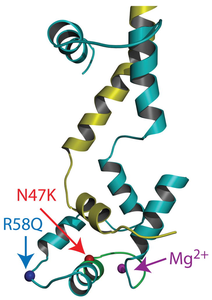
Figure 1.
FHC mutations mapped onto the chicken skeletal RLC structure (Accession # 2MYS [7]). The C-terminal region of the myosin heavy chain is shown in yellow and the RLC is shown in cyan. The locations of R58, N47, Mg2+ and the cation binding site are highlighted in blue, red, magenta and green respectively.

