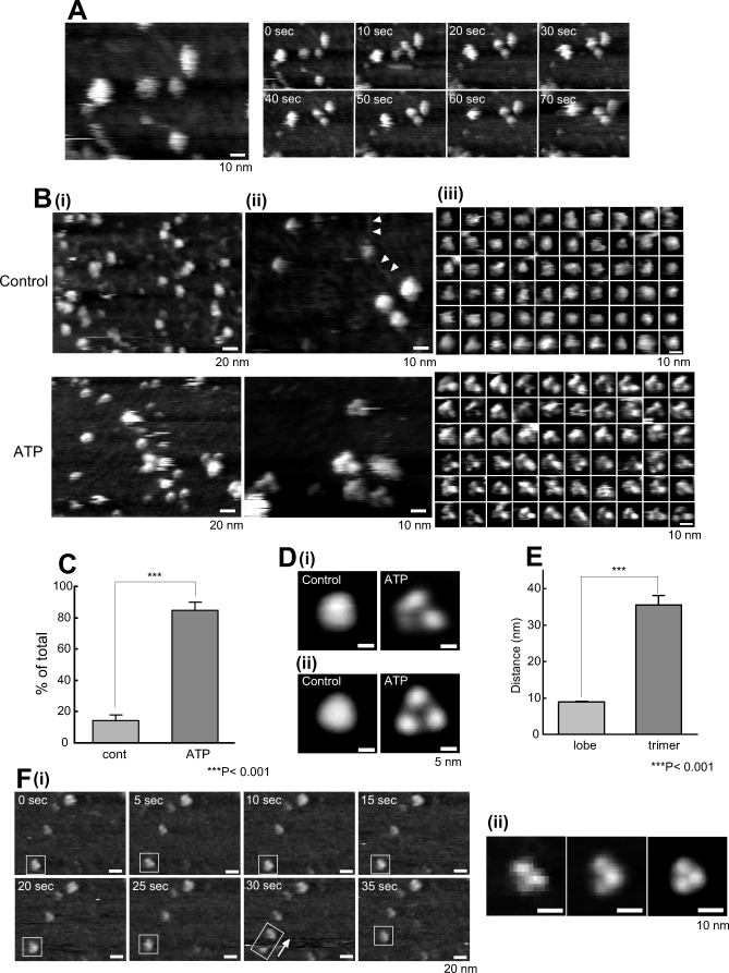
Figure 2. AFM Observations of P2X4Rs on PDL-Coated Mica.
(A) P2X4Rs attach stably to PDL-coated mica. P2X4Rs on PDL-coated mica exhibited stable attachment and the majority did not shift position during AFM observation. Scale bar, 10 nm.
(B) AFM images of P2X4Rs at (i) low resolution, (ii) high resolution, and (iii) single particle level. (i) At low resolution, the P2X4Rs were relatively homogenous. Slight differences were observed after ATP treatment (1 mM, 30 min), but they are not very clear at this resolution. Scale bar, 20 nm. (ii, iii) At high resolution and at a single particle level, there were significant structural differences between the control P2X4Rs and the P2X4Rs after ATP addition. Each single particle was selected based on our criteria (Materials and Methods, Figure S1C). In the control, the P2X4Rs were nearly circular, ellipsoid, or triangular with obtuse angles. After ATP addition, the P2X4Rs had a tripartite morphology. PDL-polymers were also observed (arrows). Scale bar, 10 nm.
(C) Percentage of trimeric P2X4R was significantly increased after ATP (1 mM, 30 min). ***, p < 0.001.
(D) Averaged images of P2X4Rs. (i) Nonsymmetrized averaging of P2X4Rs in the control (left) and after ATP addition (right). (ii) Symmetrized averaging of P2X4Rs in the control (left) and after ATP addition (right). 3-fold symmetrized images were obtained after symmetrized averaging. Scale bar, 5 nm.
(E) Three lobes are individual subunits in one P2X4R trimer. The distance between lobes was significantly less than that between trimers. ***, p < 0.001.
(F) P2X4R trimer shifts position as one unit. (i) When a P2X4R trimer moves during AFM, the three lobes were not dissociated but moved as a trimer. Scale bar, 20 nm. (ii) Enlarged images of single P2X4R trimer in a rectangle at 5 s, nonsymmetrized and symmetrized averaging images of ten particles in the same scan area. Scale bar, 10 nm. AFM observation was performed in AFM imaging buffer A.

