Figure 7.
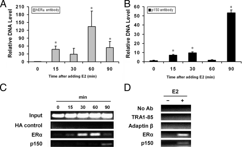
p150/glued is recruited to the estrogen-responsive pS2 promoter in MCF-7 cells. Protein association with pS2 promoter was assessed using ChIP. MCF-7 cells were plated in estrogen-free DMEM 5% fetal calf serum for 3–5 d and then pulsed with estrogen (10 nm) for periods shown. At the end of each incubation, cells were fixed with formalin and chromatin fragmented by sonication. A, ERα antibodies were used to demonstrate the temporal profile of ERα association with the ERE sites of the pS2 promoter, which was quantified using quantitative PCR (qPCR). B, p150/glued monoclonal antibodies were used to immunoprecipitate protein/DNA adducts, which were subjected to qPCR analysis using primers against the proximal pS2 promoter. The immunoprecipitated pS2 promoter DNA was normalized to the value at time zero. C and D, Endpoint PCR analysis of ChIP assay demonstrates that multiple negative control antibodies failed to precipitate pS2 target gene. Negative control antibodies, which failed to associate with the target gene (with or without estrogen), included HA, TRA1-85, and adaptin-β; in addition, reactions performed without antibody failed to precipitate detectable quantities of pS2 chromatin. This analysis is qualitative and does not precisely mirror qPCR results, which are considered more quantitative. qPCR data shown are from two independent experiments. Endpoint PCR analysis represents four independent experiments in panel C and two experiments in panel D. *, Statistical significance (P < 0.05) relative to unstimulated cells. Ab, Antibody.

