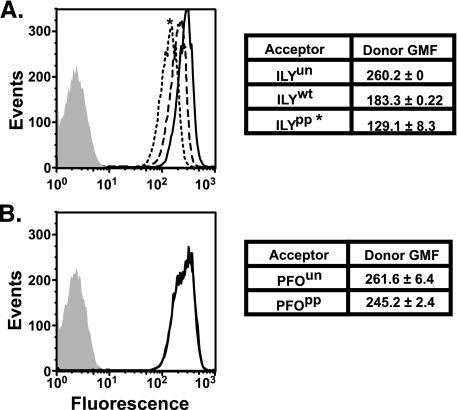
FIGURE 4.
FRET detected association between ILY and hCD59 in prepore complexes. A cysteine was introduced at amino acid residue Asp-280 in both ILYwt and ILYpp and was labeled with tetramethylrhodamine (A, acceptor fluorophore). A, erythrocytes were preincubated with a FITC-conjugated (D, donor fluorophore) anti-hCD59 mAb (MEM43) and then incubated with either unlabeled ILYun (D + U, solid line) or acceptor-labeled ILYwt (D + A, dashed line) or ILYpp (D + A, dotted line). FRET between donor and acceptor is observed as a decrease in the fluorescence per cell (i.e. the peak will shift to the left) of the donor when the unlabeled toxin is replaced with acceptor-labeled toxin (compare D + U and D + A). No significant change in donor fluorescence was observed when acceptor-labeled PFOpp (D + A, dotted) or unlabeled PFOun (D + A, solid line) was used (B). GMF, geometric mean of fluorescence. *, p < 0.005 for the geometric mean of fluorescence of cells treated with ILYpp and ILYwt. No significant difference in the geometric mean of fluorescence of cells treated with either PFOpp or PFOwt was observed.

