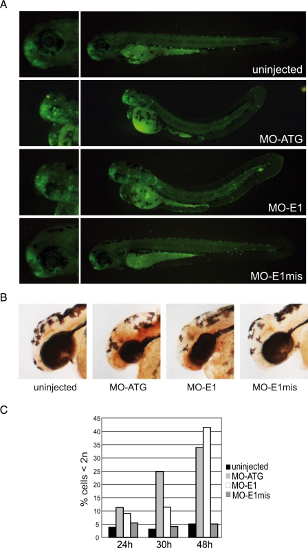
FIGURE 4.
Enhanced apoptosis in lin9 morphant brains. A, 72 hpf uninjected and morpholino (7 ng)-injected embryos were incubated with the vital dye acridine orange. Fluorescent areas are spread throughout the morphant brains, indicating abnormal apoptosis. Left, magnified views of the heads are given. B, 48 hpf uninjected and morpholino-injected embryos were paraformaldehyde-fixed, and TUNEL assays where performed. Brown 3,3′-diaminobenzidine staining indicates enhanced apoptosis in the brain and olfactory regions of lin9 morphant embryos. C, representative distribution of apoptotic cells in uninjected and morpholino-injected embryos at the indicated hpf. Cells were identified as subdiploid (<2n) by flow cytometry.

