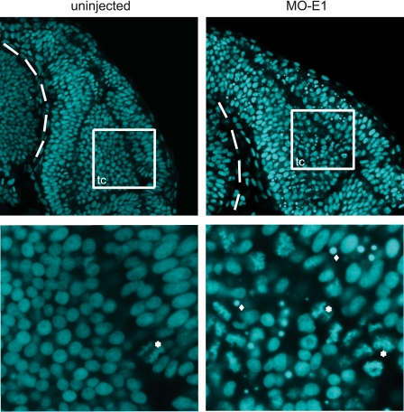
FIGURE 5.
Increase of apoptotic and mitotic cell numbers in the central nervous system of morphant embryos. Embryos were fixed and Hoechst-stained 30 hpf. Confocal images were taken of the brain (dorsal view). Dashed lines encircle the eye. Below, magnified views of wild type and E1 morphant optic tectum (tc). Examples of mitotic cells (*) and apoptotic bodies (♦) are indicated.

