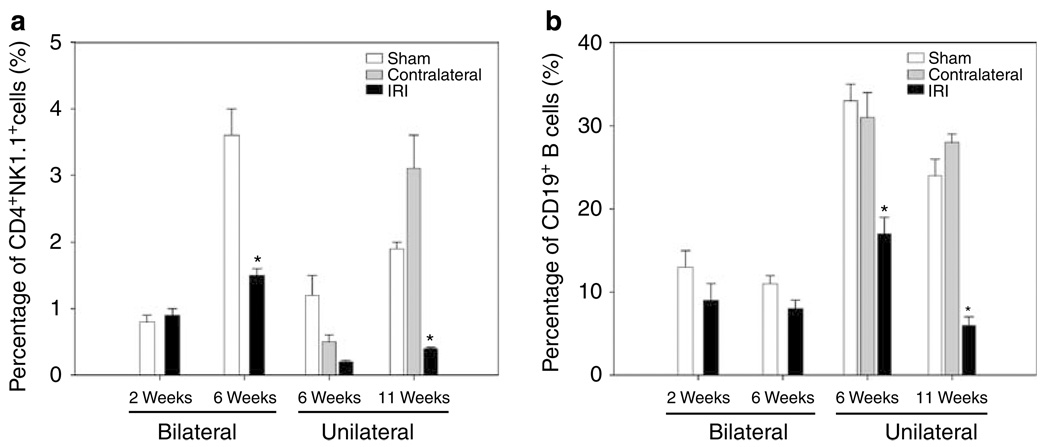
Figure 4. Decreased percentages of CD4 +NK1.1 + cells and CD19 + B cells after bilateral and unilateral renal IRI.
Decreased percentage of CD4 + NK1.1 + T cells is shown in IRI kidneys after 6 weeks of bilateral (*P = 0.007) and 11 weeks of unilateral (*P = 0.004) renal IRI when compared with control kidneys (a, left panel). The percentage of CD19 + B cells also decreased significantly in IRI kidneys after 6 (*P = 0.004) and 11 (*P < 0.001) weeks of unilateral renal IRI (b, right panel). Values of the bar graphs represent the percentage mean ± s.e.m. (n = 3 to 4 per group.)

