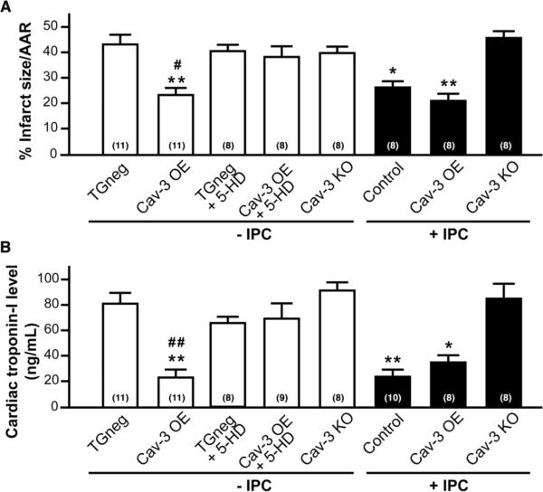
Figure 5. Cardiac protection in Cav-3 OE mice.
Mice were subjected to ischemia-reperfusion injury. (A) Infarct size (percent of AAR) was reduced by IPC in control animals; however, Cav-3 OE mice were protected to similar levels with and without IPC. Cav-3 KO mice could not be protected with IPC. Treatment of Cav-3 OE mice with 5-hydroxydecanoate (5-HD; 10 mg/kg i.v.), a mitochondrial KATP channel inhibitor, abolished protection. TGneg treated with 5-HD had similar infarct size to controls. *P < 0.05, **P < 0.001 vs. TGneg mice, and #P < 0.05 vs. Cav-3 OE + 5-HD. Group sizes are indicated on the individual bars in parentheses. (B) Serum cardiac troponin-I, a marker of cardiac myocyte damage, was measured following 2 h of reperfusion. *P < 0.05, **P < 0.001 vs. TGneg mice, and ##P < 0.01 vs. Cav-3 OE + 5-HD. Group sizes are indicated on the individual bars parentheses.

