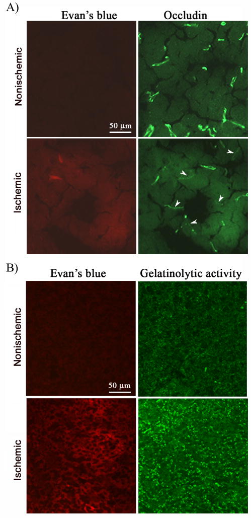
Figure 2.
Evan’s blue leakage was accompanied by occludin protein degradation and increased gelatinolytic activity in the ischemic brain of the normoxic rats after 90-min MCAO and 3-hr reperfusion. Occludin protein and gelatinolytic activity of MMP-2/9 were analyzed by immunohistochemistry and in situ zymography, respectively, on cryosections obtained from brain tissue injected with Evan’s blue. A) Immunostaining for occludin (green) was clearly seen on the microvessels of the nonischemic hemisphere, where no Evan’s leakage (red) was observed. In the ischemic hemisphere, Evan’s blue leakage was accompanied by reduced occludin staining on the microvessels. Arrowheads indicate cerebral microvessels with reduced occludin staining. Experiments were repeated three times with similar results. B) In situ zymography showed increased gelatinolytic activity of MMP-2/9 in the ischemic hemisphere (bright green fluorescence), where Evan’s blue leakage concurrently occurred. No Evan’s blue leakage and weak gelatinolytic activity were seen in the corresponding region of the nonischemic hemisphere. Experiments were repeated three times with similar results.

