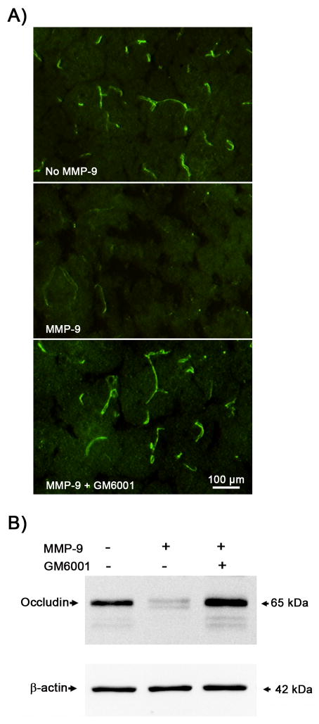
Figure 4. In vitro.
degradation of occludin protein by MMP-9. Brain cryosections or cerebral microvessel extracts obtained from normal rat brains were incubated with or without purified MMP-9 (5 μg/ml) and MMP inhibitor GM6001 (25 μmol/L) at 37°C for 2 hrs. After incubation, MMP-mediated occludin degradation in brain sections and microvessel extracts was determined by immunohistochemistry and western blot, respectively. A) The immunostaining of occludin (green) in the brain tissue almost completely disappeared after incubation with MMP-9, which was completely reversed by GM6001. Experiments were repeated three times with similar results. B) Representative western blots showed similar MMP-dependent occludin degradation in cerebral microvessel extracts. Aliquots (10 μg) of cerebral microvessel lysates were treated as indicated before they were subjected to western blot analysis. Experiments were repeated three times with similar results.

