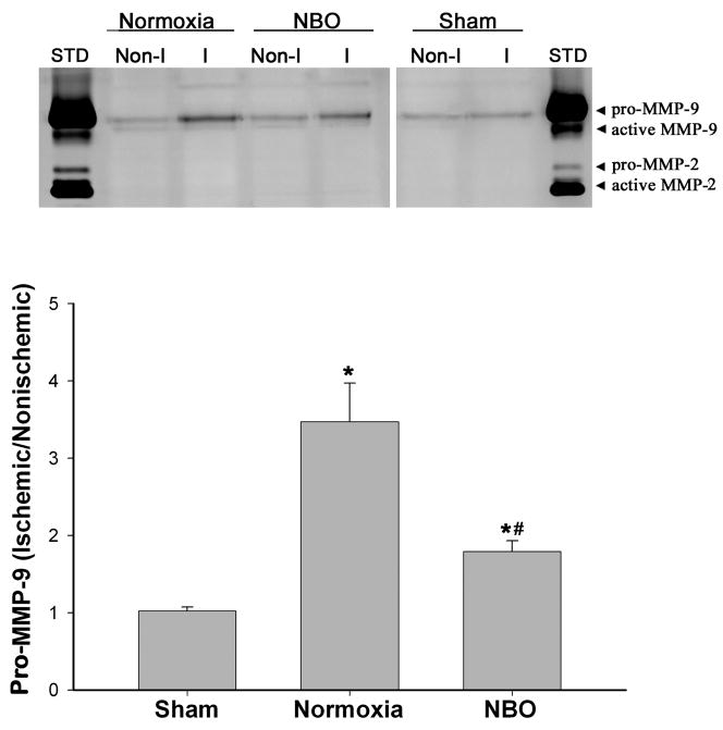
Figure 5.
NBO treatment significantly inhibited MMP-9 induction in the ischemic cerebral microvessels after 90-min MCAO with 3-hr reperfusion. Cerebral microvessels were homogenized in TCNB buffer and protein extracts (20 μg) were subjected to gel gelatin zymography analysis. Upper panel: representative gelatin zymograms showing the expression of 92 kDa proform of MMP-9 in nonischemic (Non-I) and ischemic (I) microvessels isolated from sham-operated, normoxic and NBO-treated rats. STD is a mixture of standard MMP-2/9. Lower panel: MMP-9 band intensity was quantified, and the relative quantity of MMP-9 was expressed as MMP-9 ratio (ischemic/nonischemic hemispheric microvessels). *p < 0.05 versus sham group; #p < 0.05 versus normoxic group. Data are expressed as mean ± SEM, n = 4 in the sham group, n = 6 in each of the normoxic and NBO groups.

