
Figure 4. Tumor regression is correlated with increased intratumoral effector cells and decreased immunosuppressive cells.
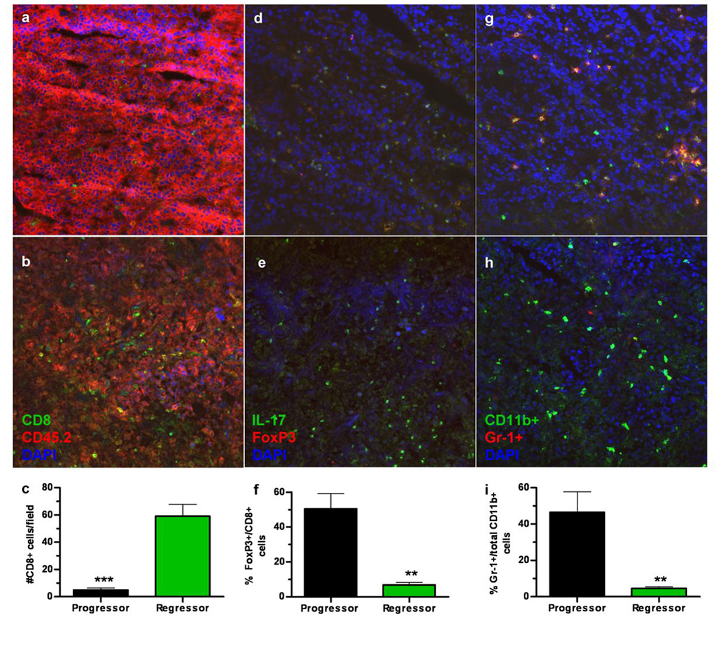
Flash frozen sections from EG7 tumors (CD45.2+) harvested from EG7-gp96-Ig vaccinated, CD45.1+ ‘progressor’ (a) mice or EG7-gp96-Ig vaccinated CD45.1+ ‘regressor’ (b) mice were stained with anti-CD8 and anti-CD45.2 (quantitated in c) or anti-IL-17 and FoxP3 (d ‘progressor’ & e ‘regressor’ and quantitated in f) or anti-CD11b and anti-Gr-1 (g ‘progressor’ & h ‘regressor’ and quantitated in i) and counterstained with DAPI (a–h). The number of positive cells was determined using ImageJ software and is plotted in panels c, f and i. ** indicates p<0.01, *** indicates p<0.001. Tumor sections were quantitated for ≥ 2 mice per group with a minimum of two sections/tumor for each staining condition with five fields per section used for counting.