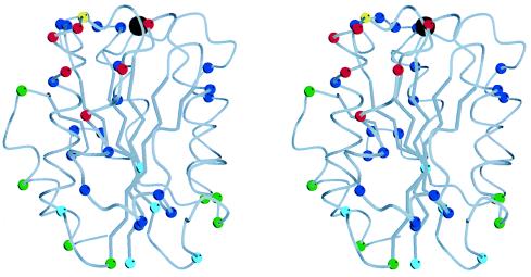
Figure 2.
Stereo view of the Mac-1 I domain (7). In the Cα trace, the Cα atoms of all residues that differ between mouse and human are shown as color-coded spheres. The black sphere at the top is the magnesium ion. The α3 helix is at front center, α-helix 1 is to its left, and the C-terminal α-helix 7 is at the far, lower left, behind α1. The Cα atoms of residues in the CBRM1/5 epitope, as defined by single amino acid substitutions, are red. The Cα of residue I146, which increases CBRM1/5 binding and decreases iC3b binding when substituted, is yellow. The remaining residues in the three human–mouse chimeras that affected CBRM1/5 binding are blue. Residues in the other 14 chimeras are color-coded according to overall effect of the substitutions: blue, decreased binding of one or more mAb and no effect on CBRM1/5 binding; green, no effect on any mAb; and turquoise, enhanced binding of CBRM1/5 mAb and no decrease in binding of any mAb. The figure was made with molscript (42).

