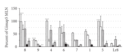
Figure 3.
Tissue specific Gimap expression. The mean ± standard deviation is shown for DR.+/+ (n = 3) Gimap gene expression. To compare Gimap gene expression across multiple tissues, data was first normalized to cyclophilin then scaled and expressed as a percentage of DR.+/+ Gimap5 mesenteric lymph node (MLN), the highest expressing gene overall. Genes appear in the order at which they appear on rat chromosome 4. Tissues appear in the following order per gene: MLN (dots), thymus (white), spleen (hash marks), bone marrow (black), and kidney (stripes). Significance is represented as follows: ∗∗∗ is P < .0001 and ∗∗ is P < .001.

