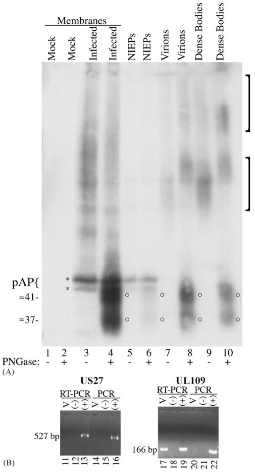
Fig. 3.
(A) N-Glycosylated GCR27 is present in HCMV extracellular enveloped particles. Cell membranes were prepared from non-infected (mock; lanes 1 and 2) or HCMV-infected (infected; lanes 3 and 4) HFF cells 8 days after infection and treated or not treated with PNGase F (±PNGase). HCMV enveloped extracellular virus particles, NIEPs (lanes 5 and 6), virions (lanes 7 and 8), and dense bodies (lanes 9 and 10), were purified from infected cells, treated or not treated with PNGase F (± PNGase), and subjected to SDS-PAGE, followed by Western immunoassay with a mixture of Anti-27 and Anti-C1, an antiserum specific for the CMV assembly protein precursor, pAP (Schenk et al., 1991; Welch et al., 1991b). Brackets denote two forms of glycosylated GCR27; circles denote deglycosylated forms of GCR27; brace and asterisks denote pAP (top) and pUL80.4, a slightly smaller protein also encoded by the HCMV UL80 nested genes (Casaday et al., 2004; Welch et al., 1991a). An image of the resulting fluorograph was captured with Adobe Photoshop for Macintosh via an Epson 1200 U scanner. (B) RT-PCR assay for UL27 and UL109 RNAs in virus particles. Clarified HCMV inoculum (100 μL) was treated with DNase-free RNase, then RNA was isolated from these samples (lanes 11, 14, 17, and 20) and subsequently treated with RNase-free DNase. Negative controls (lanes 12, 15, 18, and 21) contained no RNA; positive controls (lanes 13, 16, 19, and 22) contained approximately 50 ng of DNA from an HCMV (AD169) bacmid (Marschall et al., 2000), to show that RT-PCR and PCR did not fail because of poor enzyme cycling conditions. One-twentieth of the RNA isolated from virus inoculum was used for each experiment. Samples were amplified in the presence of an RT/Taq DNA polymerase mix (RT-PCR, lanes 11–13 and 17–19) or Platinum Taq DNA polymerase alone (PCR, lanes 14–16 and 20–22) for 60 cycles. PCR products were resolved on 2% agarose gels in SB buffer (Brody and Kern, 2004), stained with ethidium bromide, and photographed on a UV transilluminator as part of a Bio-Rad Molecular Imager Gel Doc XR system. Markers (1-kb markers or PCR markers, both from Promega) run in adjacent lanes allowed characterization of the 527-bp US27 PCR product and the 166-bp UL109 PCR product.

