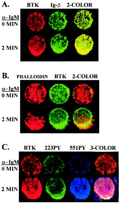
Figure 3.
Activated Btk molecules colocalize with the aggregated BCR. Cells were treated without or with anti-IgM for 2 min, fixed, permeabilized, and stained as indicated. In A, cells were stained with anti-Btk NT (secondary reagent Texas Red-labeled donkey anti-rabbit IgG) and anti-human Igβ (secondary reagent fluorescein isothiocyanate-labeled rat anti-mouse IgG1). In B, cells were stained with anti-Btk NT (secondary reagent fluorescein isothiocyanate-labeled donkey anti-rabbit IgG) and Texas Red X-labeled phalloidin. In C, cells were stained with anti-Btk NT (secondary reagent Texas Red-labeled donkey anti-rabbit IgG), 55IPYmAb (secondary reagent Cy5-labeled goat anti-mouse IgG2b), and 223PYmAb (secondary reagent fluorescein isothiocyanate-labeled rat anti-mouse IgG1). Confocal microscopic analysis was used to visualize Btk and the other proteins in representative cells.

