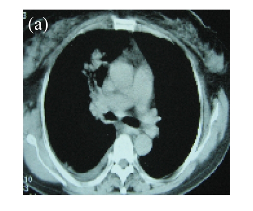
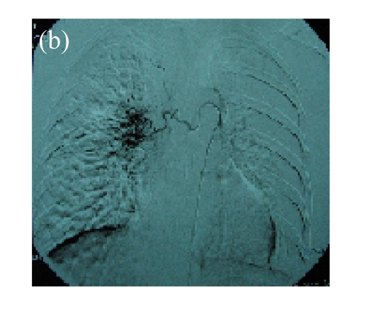
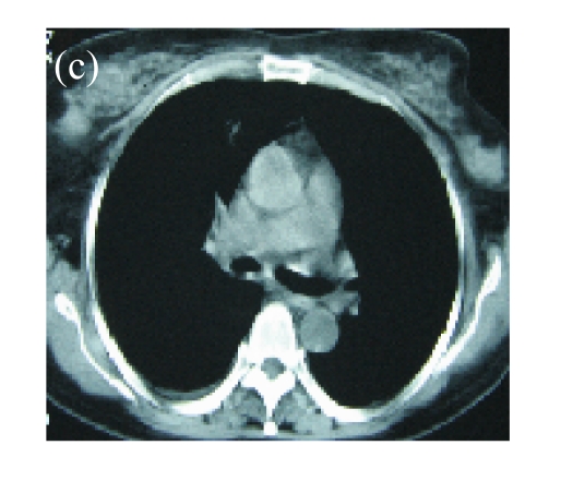
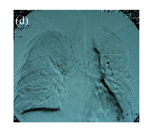
Fig. 2.
(a) A 56-year-old female with squamous cancer in the right lobe. Lesion was located in anterior segment of the right lung around the right hilus; (b) Bronchial arterial angiography showed the abnormal vascularity of tumor; (c) One month after two circles of rAd-p53 gene arterial infusion (3×10¹² VPs per time) in combination with BAI, the CT scan found that the lesions in anterior segment and around the hilus were significant regression; (d) The third BAI showed the significant reduction of tumor vascularity


