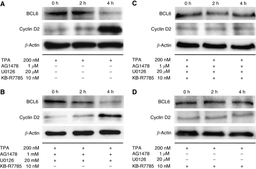
Figure 2.
Nuclear translocation of HB-EGF-CTF induces degradation of BCL6 and upregulation of cyclin D2 expression. (A) MKN45 cells were treated with 200 nM TPA for 2 or 4 h. The levels of BCL6 and cyclin D2 protein were determined by western blotting with anti-BCL6 and anti-cyclin D2 antibody. (B) Before the treatment of MKN45 cells with 200 nM TPA for 2 or 4 h, the cells were preincubated with 1 μM AG1478 and 20 μM U0126 for 60 min. The levels of BCL6 and cyclin D2 protein were analysed by western blotting. (C) Before TPA stimuli, 10 μM KB-R7785 (a metalloproteinase inhibitor) with both 1 μM AG1478 and 20 μM U0126 was added to the cells for 60 min. Then, MKN45 cells were treated with 200 nM TPA for 2 or 4 h. The levels of BCL6 and cyclin D2 protein were determined by western blotting. (D) Before TPA stimuli, 10 μM KB-R7785 alone was added to the cells for 60 min. Then, MKN45 cells were treated with 200 nM TPA for 2 or 4 h. The levels of BCL6 and cyclin D2 protein were determined by western blotting. The total incubation time from the start of the TPA treatment to cell lysis is shown at the top of the panel. β-Actin was used as a loading control for western blotting.

