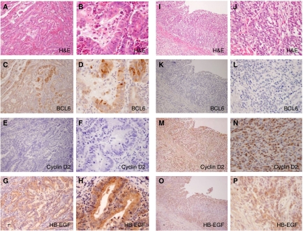
Figure 7.
Immunohistochemistry of human gastric cancers. (A–H) Differentiated gastric cancer and (I–P) undifferentiated gastric cancer. (A and B) H&E staining; (C and D) BCL6 detected in the nucleus and cytoplasm of cancer cells; (E and F) cyclin D2 was negative; (G and H) HB-EGF was detected in the cytoplasm of cancer cells; (I and J) H&E staining; (K and L) BCL6 was negative; (M and N) cyclin D2 nuclear staining is observed in cancer cells; and (O and P) HB-EGF was detected in the cytoplasm of cancer cells. (Original magnification: A, C, E, G, I, K, M, O × 100; B, D, F, H, J, L, N, P ×400).

