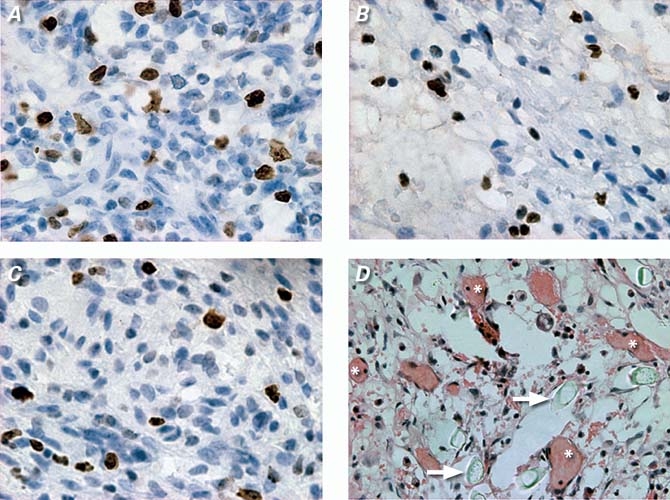
Fig. 2 Representative images obtained by Ki-67 immunohistologic and hematoxylin-eosin staining: Ki-67 positively stained S-stage cells in CFS group (A), in channels and blank scaffolds (CBS) group (B), and in control group (C) (3′3-diaminobenzidine HCl, orig. ×400). New vessels within the bFGF-incorporating scaffold (CFS) group (D) (* indicates new vessels, arrows indicate polymer remnants; H & E, orig. ×200).
