Fig. 2.
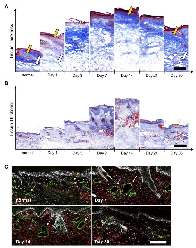
Extracellular matrix density, lipid accumulation, and macrophage presence in edematous tails. (A) In normal skin, a collagen-dense dermis (gold arrow) and less dense hypodermis (white arrow) was seen. One day following lymphatic ligation, the collagen network was degraded in both the dermis and hypodermis as fluid accumulated. At day 14 when tail volume reaches its peak, the collagen density (blue) in the dermis returned to normal (gold arrow) whereas the architecture of the hypodermis remains severely compromised. At day 30, the collagen architecture throughout the tissue appeared normal. (B) Oil red O staining showed that the hypodermis dramatically filled with lipids (red) at day 7 and that this accumulation was maintained through 30days. (C) Macrophages (red) were also present in high numbers at all times during edema. Note the hyperplastic lymphatic (green) vessels at days 7 and 14 (marked by white arrows) as compared to normal vessels (gold arrows). Scale bars=200 μm. (For interpretation of the references to colour in this figure legend, the reader is referred to the web version of this article.)

