
Figure 6.
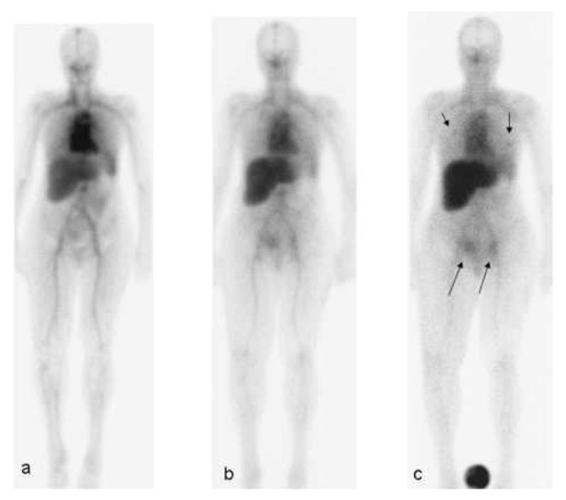
Patient with metastatic ovarian cancer. The patient underwent imaging after intravenous injection of 7.22 mCi of 111In DOTA MORAb-003, co-infused with a total of 677 mg of MORAb-003 (400 mg/m2). Serial 23 minute whole body scans were obtained within 2 hr of injection (a), 2 days (b) and 5 days (c). At 5 days a standard is shown in the field of view between the patient's feet. Arrows indicated areas of metastatic ovarian cancer visualized, 1 in each lung (short arrows) and in the right anterior pelvic wall lesion and a left inguinal node (long arrows).