Figure 1.

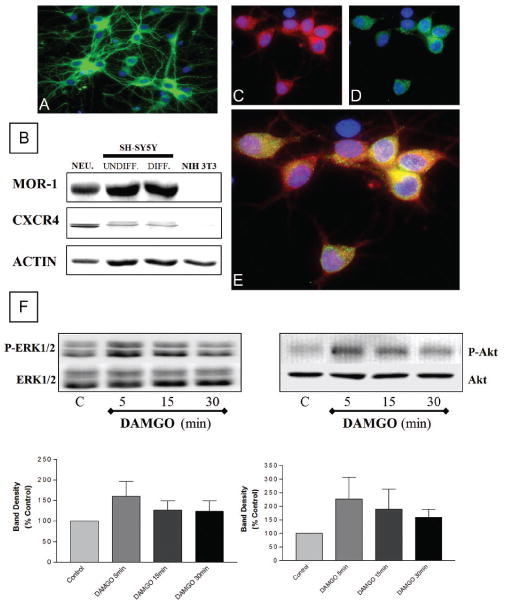
Coexpression of MOR and CXCR4 in cortical neurons. Expression of β-tubulin III (A, green), MOR (C, red), and CXCR4 (D, green) in cortical neurons was detected by immunocytochemistry and fluor escence microscopy. Nuclei are stained with Hoechst 33342 (A-E, blue). Double staining (E) shows coexpression of MOR and CXCR4 on individual neurons. Immunoblots (B) confirm presence of both receptors in cortical neurons. SH-SY5Y and NIH-3T3 cells were used as positive and negative control, respectively; actin levels were determined to verify protein loading. DAMGO (10 μM, 5–30 min) increased phosphorylation of ERK1/2 and Akt in neuronal extracts (F). Graph shows average data from four experiments (top: phosphorylated ERK1/2 [P-ERK] or Akt [P-Akt]; bottom: total ERK1/2 or Akt). However, the kinetics of response for both ERK and Akt is not identical among different experiments (which is expected in primary cultures) and this reflected by the data in the graph showing mean ± SEM for ERK (n = 3) and Akt (n = 4), respectively. Thus, for statistical analysis purposes, peak responses from these experiments (5′ and 15′) were combined and compared to controls (P < .05).
