Figure 4.

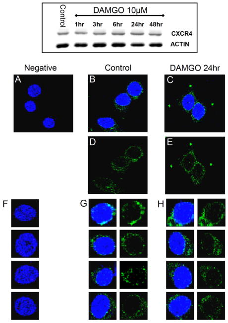
Effect of DAMGO on total and plasma membrane levels of CXCR4 in cortical neurons. CXCR4 protein levels in cortical neurons were studied by Western blot (top panel) and confocal microscopy (A–H) as described in Methods. As shown by the representative gel in the top panel (n = 3), cortical neurons were treated with DAMGO up to 48 h. The confocal microscopy studies were preformed on cultures treated with DAMGO (1 μM) for 24 h. In both sets of experiments, control and DAMGO-treated neurons appeared to have similar levels of CXCR4. For the confocal studies, neurons were stained with an antibody against CXCR4 (green) and the nuclear dye Hoechst 33342 (blue) as detailed in methods section, and visualized using a 63X 1.4 oil immersion objective mounted on a Leica confocal microscope. Images were collected by using a step size of 0.3 μm along the z axis (512 × 512; 8 bits/pixel). A and F: Negative control (i.e., without primary antibody). B, D, and G: Control. C, E, and H: DAMGO-treated. A total of 100 cells per treatment were studied from two independent experiments (three coverslips/group).
