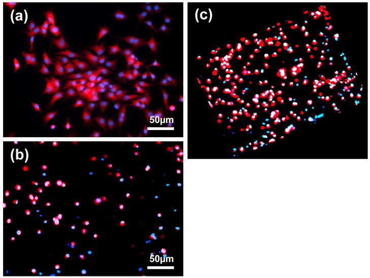
Figure 9.
(a) CLSM image showing bovine chondrocytes upon the surface of a 5/5 S-CS/A-HA composite hydrogel after 24h culture. Cell seeding density: 50,000/well (24-well cell culture plate). (b) CLSM image showing bovine chondrocytes encapsulated in a 5/5 S-CS/A-HA composite hydrogel after 24h culture. Cell seeding density: 5×10⁶/mL. (c) The 3D image of (b). The live cells were dyed with Cell Tracker Orange CMRA (Red) and all cell nuclei were dyed by Hoechst 33342 (Blue).

