Figure 1.
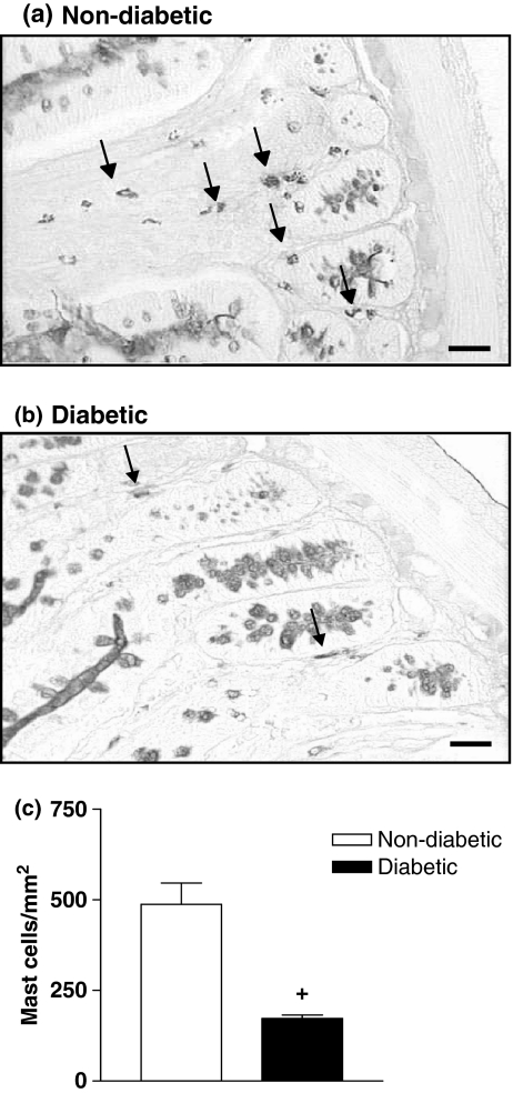
Decrease in mast cell numbers in the intestine of diabetic rats. Panels (a) and (b) show photomicrographs of PAS-alcian blue/safranin stained mast cells (arrows) from ileum segments of non-diabetic and alloxan-diabetic rats respectively. Bar 0.1 μm. Panel (c) shows that the number of mast cells is significantly reduced in the intestine of diabetic rats compared with controls. Data are expressed as mean ± SEM from three animals. The data are representative of three experiments. +P < 0.05 compared with non-diabetic rats.

