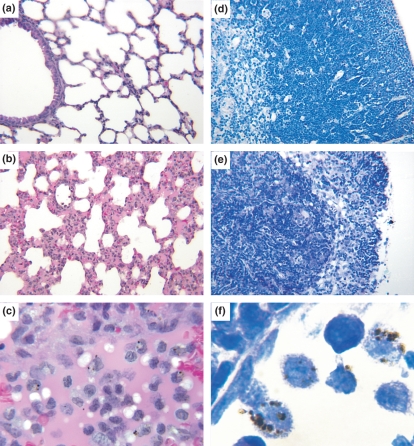
Figure 3.
Lungs and thymus of Swiss Webster mice. (a) Panoramic view of non-infected control mouse lung with thin alveoli walls and clean blood vessels. Haematoxylin–eosin, 200×. (b) Panoramic view of the lung of an infected mouse, showing thickening of alveoli walls by the infiltration of mononuclear and polymorphonuclear cells. Haematoxylin–eosin, 200×. (c) Detail of septal thickening in an infected mouse lung, showing predominance of mononuclear cells. Haematoxylin–eosin, 400×. (d) Panoramic view of a normal mouse thymus, showing the cortical area plenty of thymocytes and the internal medullar area with a lower cellularity. Giemsa, 200×. (e) Panoramic view of the thymus of a infected mouse, showing intense thymocyte depletion in the cortical area, with only a few remaining thymocytes between the framework of epithelial cells, and an increase in the medullar cellularity resulting in cortico-medullar inversion. Giemsa, 200×. (f) Pronounced monocyte adherence on the endothelium of thymus medullary vessels of infected mice. Monocytes look like macrophages and contain malarial pigment. Giemsa, 1000×.

