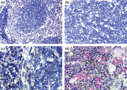
Figure 4.
Spleen of Swiss Webster mice. (a) Panoramic view of non-infected control mouse spleen. White and red pulps with well-defined limits. One B-cell resting follicle with surrounding thick marginal zones. Giemsa, 200×. (b) Panoramic view of infected mouse spleen. Disorganized germinal centre with intense centroblast activation, proliferation, apoptosis, without centrocyte differentiation and definition of light and dark areas, absence of marginal zone and blurred limits between white and red pulps. Giemsa, 200×. (c) T-cell area (periarteriolar lymphoid sheath) of infected mouse. Centroblasts. Giemsa, 400×. (d) Infected mouse, malarial pigment in red pulp macrophages. Giemsa 400×.

