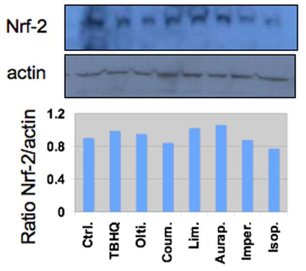
Figure 2.
Western blot analysis of Nrf2 protein in HepG2 cells. HepG2 cells were treated with vehicle (control, ctrl., DMSO 0.1% v/v), or 25 μM of tBHQ, oltipraz (olti.), coumarin (coum.), limettin (lim.), auraptene (aurap.), imperatorin (imper.) and isopimpinellin (isop.) for 24 h. Whole cell lysates were immunoblotted for Nrf2 and compared to β-actin as a loading control. The lower panel shows the ratio of the integrated density of Nrf2:actin.

