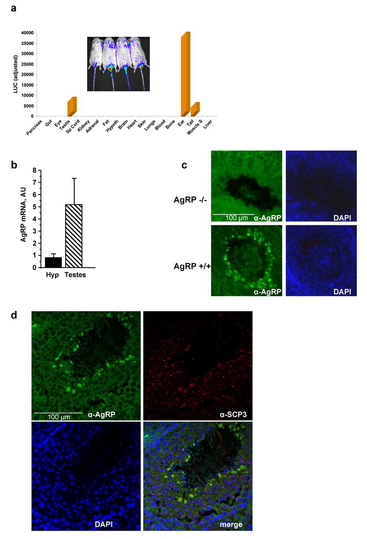
Fig. 4. Luminometric analysis of luciferase signal in Diiia mice and localization of AgRP by immunofluorescence.

(a) Luminometric analysis (light units) of luciferase expression in various tissues of Diiia mice (inset shows bioluminescence of a ventral view of these mice). Luciferase is predominantly expressed in the ears, testes, and tails. (b) Real time PCR shows robust expression of AgRP in the testis in comparison to AgRP in the hypothalamus. (c) Immunohistochemistry of mouse testis sections localized AgRP in the middle of seminiferous tubules in wild type (AgRP+/+) but not AgRP-deficient (AgRP-/-) mice. (d) Immunohistochemistry colocalized AgRP with the alpha-synaptonemal complex protein 3 (α-SCP3) in pachytene stage spermatocytes.
