
Fig. 5. Luminometric analysis of luciferase signal in Diiib mice and localization of AgRP by immunofluorescence.
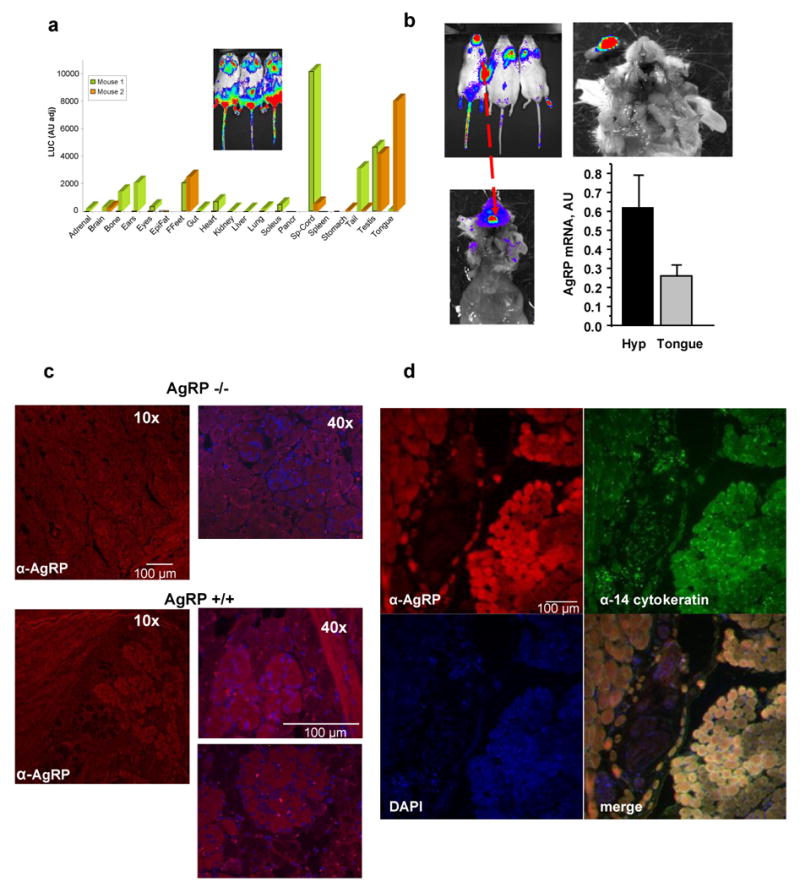
(a) Luminometric analysis (light units) of luciferase expression in various tissues in Diiib mice (inset shows bioluminescence of a ventral view of these mice). Luciferase is ubiquitously expressed with greater preference for the spinal cord, the tongue, the testis, the ears, the bone, the brain, front feet, soleus muscle, among other tissues. There was significant variation between the two founders indicated by different colors of the bars. (b) Bioluminescence of the signal in the tongue is shown it different zoom modes. Real time PCR shows robust expression of AgRP in the tongue but at lower levels than in the hypothalamus. (c) Immunohistochemistry of mouse tongue sections shows AgRP expression in epithelial cells of wild type (AgRP+/+) but not AgRP-deficient (AgRP-/-) mice. (d) Immunohistochemistry colocalized AgRP with the α-14-cytokeratin that is an epithelial cell-specific protein.