
FIGURE 1.
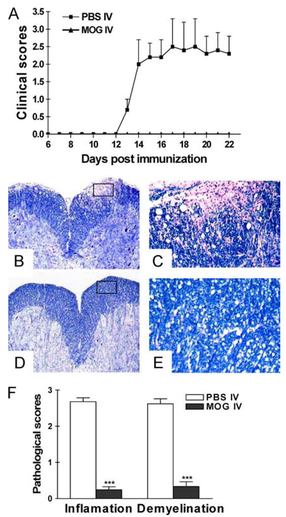
Intravenous injection of MOG35–55 prevents EAE. C57BL/6 mice were immunized with MOG35–55 + CFA. Pertussis toxin was injected on days 0 and 2 p.i. Two hundred micrograms of MOG35–55 was injected i.v. on days 3, 5, and 7 p.i. Mice that received i.v. PBS in parallel served as controls. Clinical EAE was scored according to 0–5 scale (A) (n = 5 in each group). The differences between PBS-i.v. mice and all MOG-i.v. groups were significant (p < 0.01). On day 21 p.i., spinal cords were harvested after extensive perfusion, and 5-μm sections were stained with H&E and Luxol fast blue. Shown are examples of Luxol fast blue staining for mice that received i.v. PBS (B and C) and i.v. MOG (D and E). B and D, Magnifications, ×10. C and E, Magnifications, ×20. The difference between PBS-i.v. and MOG-i.v. mice was significant (p < 0.01). F, Mean scores of inflammation and demyelination ± SD in MOG-i.v. and PBS-i.v. mice (n = 5 each group). ***, p < 0.001. One representative experiment of three is shown (total n = 15 in each group).