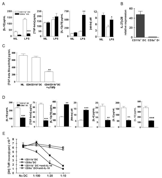
FIGURE 5.
Characteristics of CD11c+CD11b+ and CD11c+CD8α+ DC subsets. A, Freshly prepared CD11c+CD11b+ or CD11c+CD8α+ DCs from splenocytes of tolerized mice were stimulated with 1 μg/ml LPS for 24 h. Supernatants were assayed for IL-10, TGF-β, IL-12p70 by ELISA, and NO by Griess assay. B, CD11c+CD11b+ and CD11c+CD8α+ DCs were purified from splenocytes of tolerized mice and IL-27 mRNA expression was determined using real-time PCR. The y-axis represents the fold increase in IL-27p28 mRNA expression in CD11c+CD11b+ when compared with CD11c+CD8α+ DCs. C, TGF-β bioactivity in the CD11c+CD11b+ cell culture supernatants was determined as described in Materials and Methods. Neutralizing Abs against TGF-β1, β2, and β3 (5 ng/ml each) were added in parallel. D, MOG-reactive CD4+ T cells (1 × 10⁵) from mice with EAE were cocultured with 1 × 10⁴ CD11c+CD11b+ DCs or CD11c+CD8α+ DCs from tolerized mice for 3 days in the presence of MOG35–55 (10 μg/ml). Concentrations of IL-10, IL-12, TGF-β1, IL-4, IL-5, and IL-13 in the culture supernatants were determined by sandwich ELISA and NO by Griess assay.*, p < 0.05;**, p < 0.01;***, p < 0.001. One representative of three experiments is shown. E, Freshly prepared CD11c+CD11b+ or CD11c+CD8α+ DCs were cocultured with MOG-reactive CD4+ T cells (1 × 10⁵) and irradiated APCs (Irrad. APC; 1 × 10⁵) obtained from the spleen of mice with EAE, for 3 days at various DC:CD4+T cell ratios in the presence of MOG35–55 (10 μg/ml). Proliferative responses of CD4+ T cells were determined. Neutralizing anti-IL-10 mAb (5 μg/ml) was added in parallel in indicated wells. Values are the mean and SD from 3 independent experiments.

