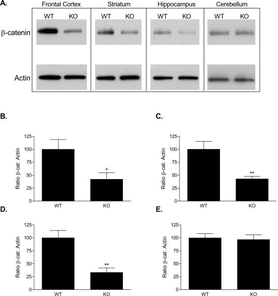
Figure 3.
β-catenin protein expression in knock-out mice at 16-20 weeks. Western blot revealed decreased expression in the frontal cortex, striatum, and hippocampus, but no difference in the cerebellum (A). Densitometry revealed that β-catenin protein levels were reduced by 58% in the frontal cortex (B), 57% in the striatum (C), and 67% in the hippocampus (D). No difference was observed in the cerebellum (E).

