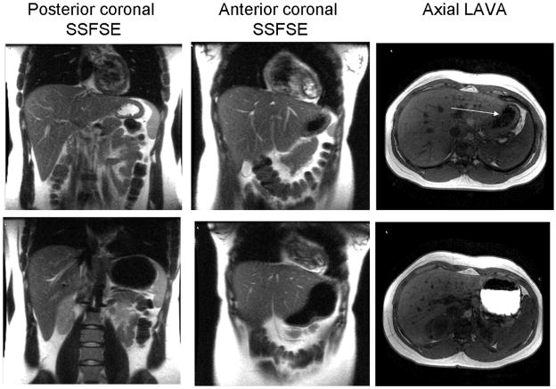
Figure 2.
Fasting (upper panel) and postprandial (lower panel) images acquired by MRI. In the fasting HASTE images, the distinction between high signal intensity fluid, which is layered posteriorly (i.e., in the dependent position) and low signal intensity air, which is anterior, is obvious. In the LAVA images, the air-fluid interface is subtle (arrow). Postprandially, air and fluid have low signal intensity in HASTE images but easily distinguishable on LAVA sequence (i.e., nutrient labeled with gadolinium appears bright while air is dark)

