
Figure 4. XY chromosome fluorescence in situ hybridization (FISH).
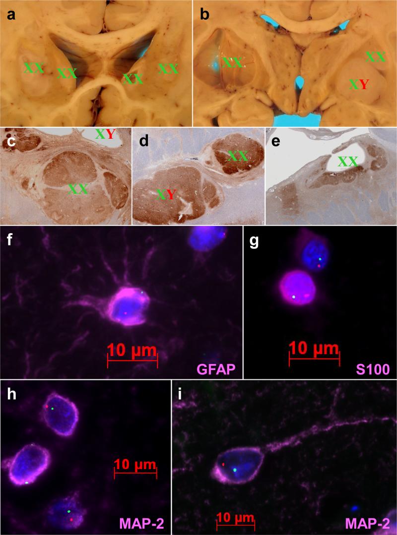
A-E) Gross (A-B) and microscopic (C-E) depictions of graft chromosomal identity, wherein graft masses are labeled with XX for female and XY for male. Cysts are labeled for XX/XY signal in ependyma lining cyst walls (B,C,E). Microscopic sections (C-E) are stained with antibodies to MAP-2 in order to highlight the grafts. F-G) High magnification fluorescence photomicrographs showing astrocytic (F), oligodendroglial (G), and neuronal (H-I) differentiation of XY chromosome-positive cells. X chromosomes are stained green and Y chromosomes are stained red. Immunostains are colored pink, and are labeled accordingly. Note the stellate processes in the GFAP immunopositive astrocyte, variable nuclear immunopositivity in small round nuclei characteristic of oligodendroglia, and the immature (H) and mature (I) morphology of MAP-2 immunopositive graft neurons.