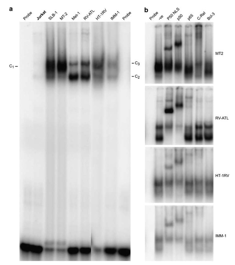
Figure 3.
NF-κB binding activity on a consensus κB binding site in various cell lines. (a) Approximately 5 µg of nuclear lysates from the cells were incubated with a [32P]-radiolabeled oligonucleotide corresponding to MHC class 1 gene κB sequence and analyzed by EMSA. The complexes are indicated as C1, C2 and C3. (b) In supershift assays, antibodies specific for each NF-κB subunit (indicated above the lane) were incubated with the nuclear extracts from MT-2, RV-ATL, HT-1RV and IMM-1 cells before addition of the probe.

