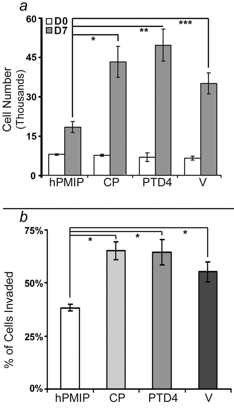
Figure 3. PMIP inhibits cell proliferation and invasion of breast cancer cells in vitro.
a) BT20 cells were cultured in a 96-well dish (5 × 10³ cells/well) and treated daily for 7 days with either hPMIP (10µM), CP (10µM), PTD4 (10µM) or water (V) in EMEM 10% FBS. An MTT assay was performed to quantify cell number at the start of treatment (Day 0, DO) and after treatment was complete (Day 7, D7) (*, p<0.001, **, p<0.0002, ***, p<0.002, ANOVA). Error bars represent standard error. b) BT-20 cell lines were treated with either hPMIP (10µM), CP (10µM), PTD4 (10µM) or water (V) overnight, labeled with Calcein AM, allowed to invade through a transwell (8.0µM) insert into a Type I collagen gel, and the invaded cells were fluorescently measured. (#p<0.0001, ANOVA). The data from the invasion and proliferation assays represents four independent experiments with at least seven replicates per experiment. Error bars represent standard deviation.

