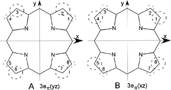
Figure 3.

Symmetry-adapted substrate 3eπ molecular orbitals that illustrate the position of significant delocalized π-spin density on the substrate resulting from singly-occupied metal-porphyrin π-bonding. The magnitude of the spin density, ρπ , is shown by the size of the dashed circle. (A) 3eπ(yz), which interacts only with dyz and results in large low-field methyl contact shifts for positions 2, 3, 6 and 7, as observed (18) for the cyanide complexes of α-meso selective hHO, and (B) 3eπ(xz), which interacts only with dxz and results in large low-field methyl contact shifts at positions 1, 4, 5 and 8, as observed in the azide complexes of α-meso selective hHO. The orbitals in (A) and (B) are similarly occupied in all α-meso selective HO cyanide (18, 19, 22, 40, 41, 46) and azide (34, 35) complexes, respectively.
