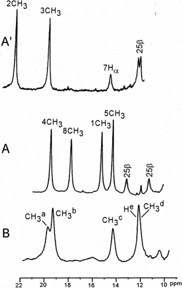
Figure 4.

Resolved portions of the 600 MHz 1H NMR spectra of: (A) hHO-DMDH-N3; (B) D140A-hHO-DMDH-N3; and (A') Reproduces the same spectral portions for hHO-DMDH-CN (18). All samples are at 30°C in 2H2O, 50 mM in phosphate at pH 7.4. DMDH peaks are labeled by the Fisher notation (see Figure 3), and residue peaks are labeled by the residue number and its position within the residue.
