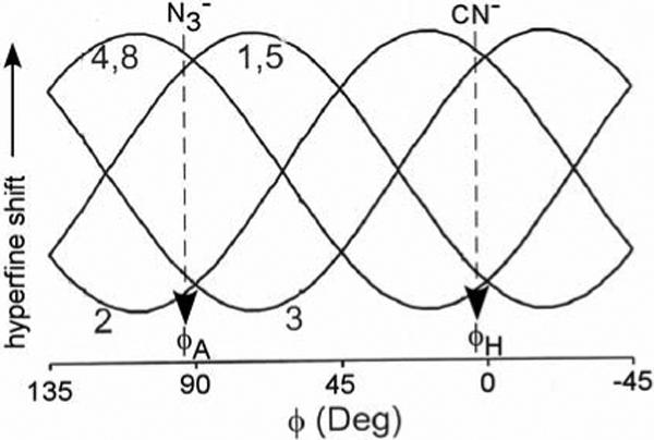
Figure 8.

Empirical plot of DMDH methyl shifts as a function of the angle φ between the unique π-plane of the axial ligand that serves as the stronger π-donor to the iron in a S = 1/2, (dxy)2(dxz,dyz)3 ferrihemoprotein. (27, 44) The angle, φ, is defined in Figure 2A. The pattern of the observed DMDH methyl shifts, and the corresponding φ, are indicated by vertical arrows under CN− (for the cyanide complex) and under (for the azide complex).
