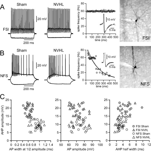
Figure 3.
Basic membrane properties of PFC interneurons in NVHL and sham rats. A, Traces illustrating the firing pattern of FSIs in PFC slices from developmentally mature (older than P60) sham and NVHL rats. FSIs exhibited a pronounced fast AHP (middle panel inset) and responded with a constant instantaneous firing rate throughout the current pulse (middle panel: no spike-frequency adaptation). Right, Neurobiotin staining of the FSIs from which the traces were obtained. B, Traces from an NFS interneuron, which instead show increasingly longer interspike intervals when activated with current injection (left and middle panels). Right, Neurobiotin staining of the interneuron recorded. C, Plots of AHP amplitude, half-width, and action potential amplitudes and duration for both cell types.

