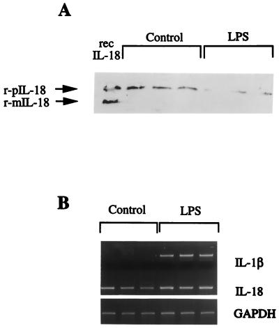
Figure 5.
Differential expression of IL-18 and IL-1β in mouse spleen. Spleens from three C57BL6 mice were removed. (A) IL-18 as assessed by Western blotting. The molecular mass markers (in kDa) of murine proIL-18 (pIL-18) or mature (mIL-18) are shown. Under the bracket labeled control, each lane represents spleen cells from a different mouse. Under the bracket labeled LPS, the mice were injected 2 h previously with LPS (100 μg). (B) Gene expression for IL-1β and IL-18 under conditions identical to those in A.

