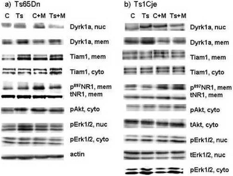
Figure 4. Sample Western blots, chr21 and non-chr21 proteins in cortex.
Approximately 30 ug of each protein lysate were resolved on 12% polyacrylamide gels (Erk1/2, Akt, Elk) or 8% or 10% gels (Dyrk1a, Tiam1) and transferred to PVDF membranes. In addition to the antibodies for the indicated proteins, each membrane was simultaneously or sequentially probed with an antibody for actin. Due to variations in the amounts of protein loaded, visual interpretation of signal intensities is not accurate and actin signals were used for normalization to correct for variable protein levels; only one example of a set of actin signals is shown. C, euploid control sample; Ts, trisomic sample; C+M, euploid control injected with MK-801; Ts+M, trisomy injected with MK-801. Nuc, nuclear; cyto, cytoplasmic; mem, crude membrane fraction. p, phosphorylated protein; t, total protein, phosphorylated plus non-phosphorylated.

