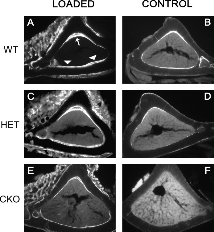
FIG. 1.
Attenuated mechanical stimulation of new bone formation in mice with conditional ablation of Gja1. Fluorescence micrographs of calcein-labeled tibial mid-diaphysis cross-sections corresponding to the area subjected to mechanical load (A, C, and E) and to the same area in unloaded, contralateral tibias (B, D, and F). Note also the thinner tibial cortex and larger marrow area of the CKO mice (E and F) compared with that of the WT (A and B). (A) Double calcein labels are present in the endocortical surface of loaded bones of wildtype Gja1+/flox mice (WT) at the apex (large arrow) and on the opposite endocortical surface (arrowheads), corresponding to the areas of maximum endocortical compression and tension, respectively. (C) Double-labeled surfaces are also seen in Gja1–/flox tibias (HET), whereas (E) predominantly single labels are present in ColCre;Gja1–/flox (CKO) tibial sections. (B, D, and F) Primarily single labels are seen in control, unloaded bones. Note also the periosteal woven bone around the area of loading, evident for each genotype.

