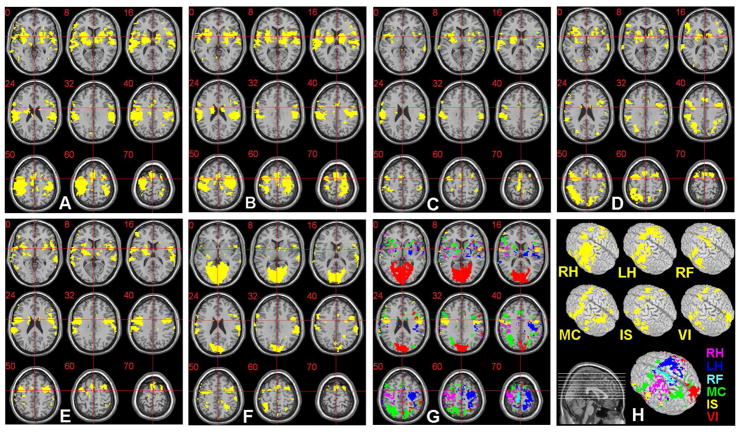
Figure 3.
The ROIs (p<0.01; z-score>2.58; d.f.=49) obtained from the six imagery tasks performed by a male subject (S4) are shown as yellow-colored regions: (A) ROI for the right-hand motor imagery (RH), (B) left-hand motor imagery (LH), (C) right-foot motor imagery (RF), (D) mental calculation (MC), (E) internal speech generation (IS), and (F) visual imagery (VI). (G) The exclusively activated regions across (A) ~ (F) are displayed in different colors (labels in H) and are overlaid on the anatomical template (provided by MRIcro; www.mricro.com v.1.39) after the normalization process. Slice range: Z=0 to +70mm (Talairach coordinates). The voxels within these exclusive ROIs were utilized as elements of a feature vector necessary for the SVM classification. (H) The original ROIs and exclusive ROI, rendered on a 3-D volume, are shown.

