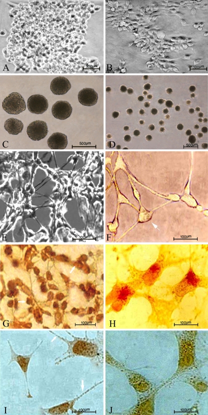
Fig. 1.
Sigmacote—coated glass petri dishes were used to form EBs from murine ES (CCE line) cells and EC (P19 line) cells. The EBs that formed differentiated into neural cells. Photomicrographs a and b show undifferentiated CCE and P19 stem cells, respectively, which were cultured as monolayer. Photomicrographs c and d show EBs derived from CCE and P19 cells, respectively, after culturing the cells for 2 days on Sigmacote™ as a substrate. EBs derived from P19 cells were cultivated from day 0 to day 4 in the presence of 0.5 μM RA. EBs derived from CCE cells were cultivated from day 0 to day 2 in the medium without RA, then on days 3 and 4 in the presence of 0.5 μM RA as the agent to induce neural differentiation. Neuron-like cells derived from CCE cells (e) and P19 cells (f) exhibited many cytoplasmic extensions from the cell body and formed synapse-like connections with the other cells. The photomicrographs g and h show DAB-immunostaining of differentiated EBs derived from CCE and P19 cells. Neural progenitor nestin positive cells derived from CCE and P19 cells, respectively. Photomicrographs i and j show synaptophysin positive neural cells differentiated from CCE and P19 cells, respectively. Magnification of d is 100×, other magnifications are 200×

