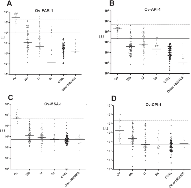
Figure 2. LIPS detection of antibodies to 4 different Ov antigens.
Each symbol represents individual samples from the 38 Ov-infected, 90 Wb, 90 Ll, 27 Ss, 72 control uninfected samples and 12 other control patients. Antibody levels in LU are plotted on the Y-axis using a log10 scale and short solid horizontal lines indicate the geometric mean titer (GMT) for each antibody per group. The diagnostic performance related to cross-reactivity with other filarial infections was also evaluated. As described in the text, the long solid line represents the cut-off level corresponding to 100% sensitivity, while the long stippled line corresponds to the cut-off for 100% specificity with sera from the Wb cohort.

